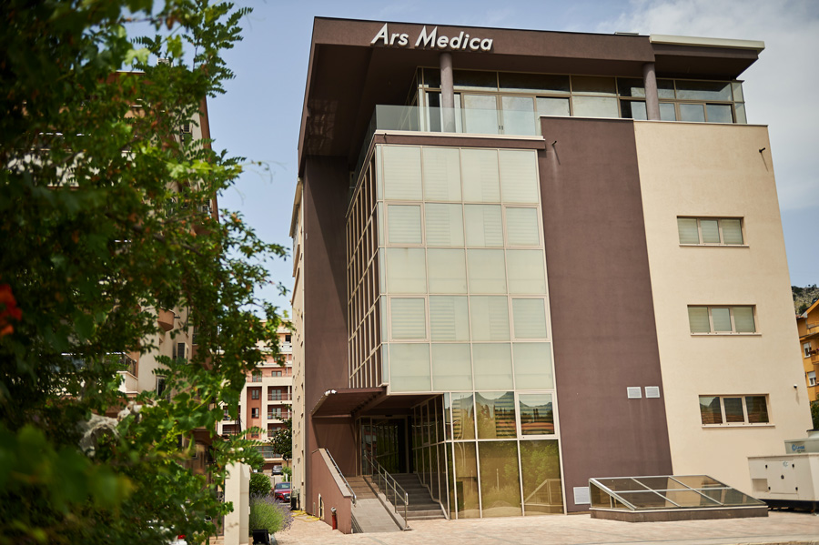
Contact
Contact
Gavra Vukovića b.b, Podgorica
Dentistry: 020 227 227
Polyclinic: 020 227 224
Gynecology: 020 227 444
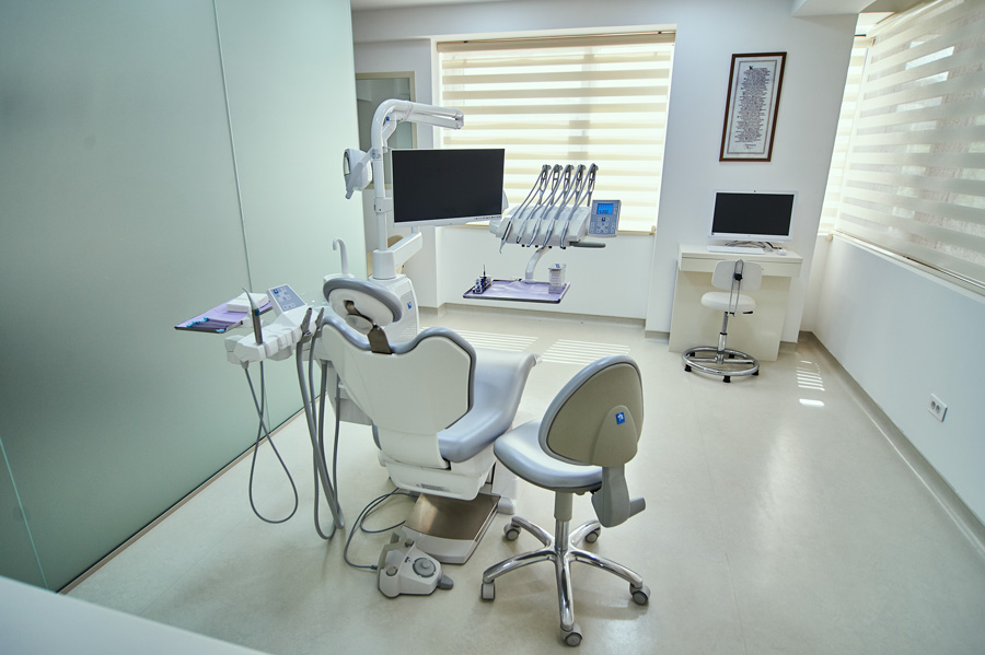
DENTISTRY
High quality dental care. Years of experience in creating perfect smiles


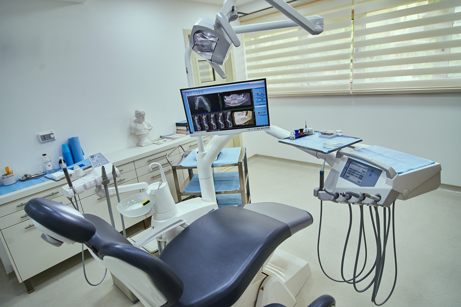
DIAGNOSTICS
Healthcare, wellbeing , aesthetics
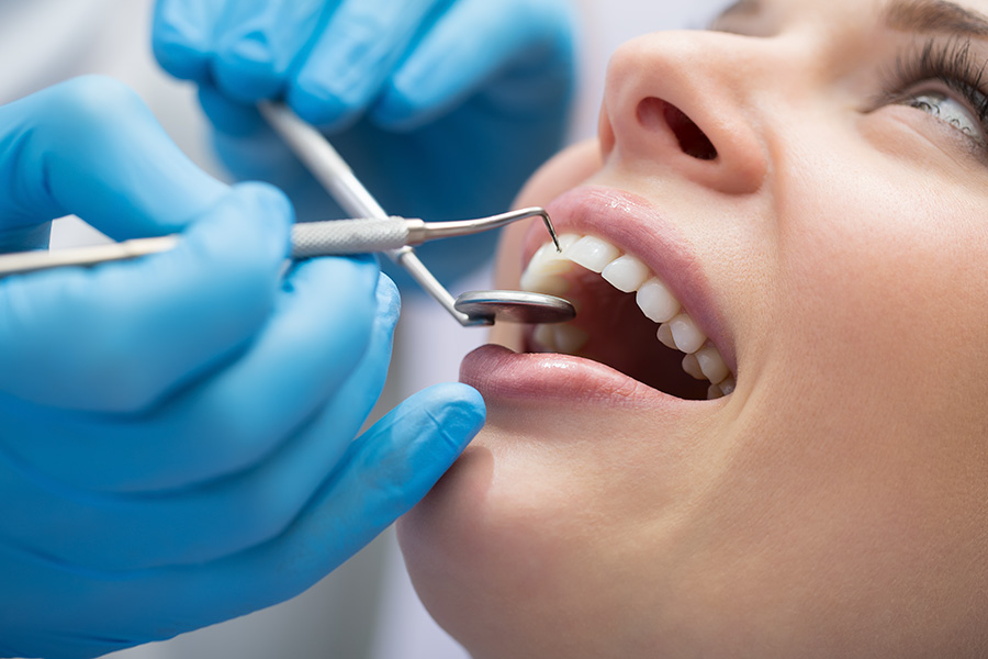
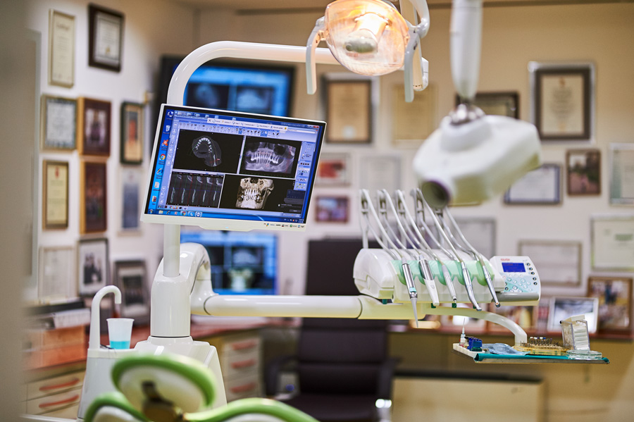
Comprehensive and precise diagnosis is the most important prerequisite for a successful treatment. Our dental clinic contains all available cutting edge diagnostic tools required for precise treatment planning concerning the pathologies of oral cavity.
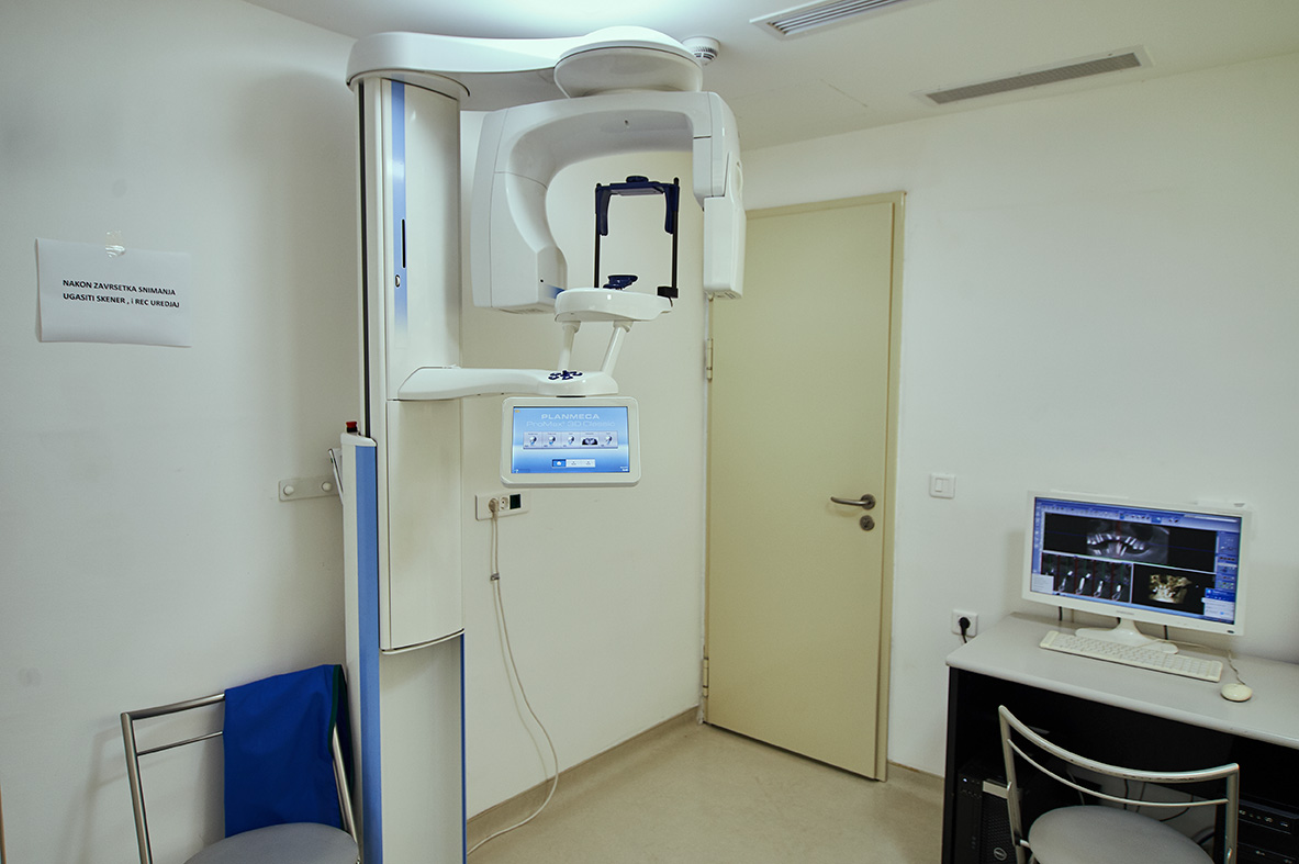
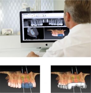
3D CBCT Scanner Planmeca ProMax
This kind of dental scan is useful for treatments in implantology, dental surgery, endodontics, periodontology and prosthodontics, due to its ability to process data in all three dimensions, thus contributing to the successful treatment execution. The process of preforming a scan is quick and painless. One scan has all the necessary information for planning a comprehensive rehabilitation of oral cavity.
3D CBCT Scanner Planmeca Promed
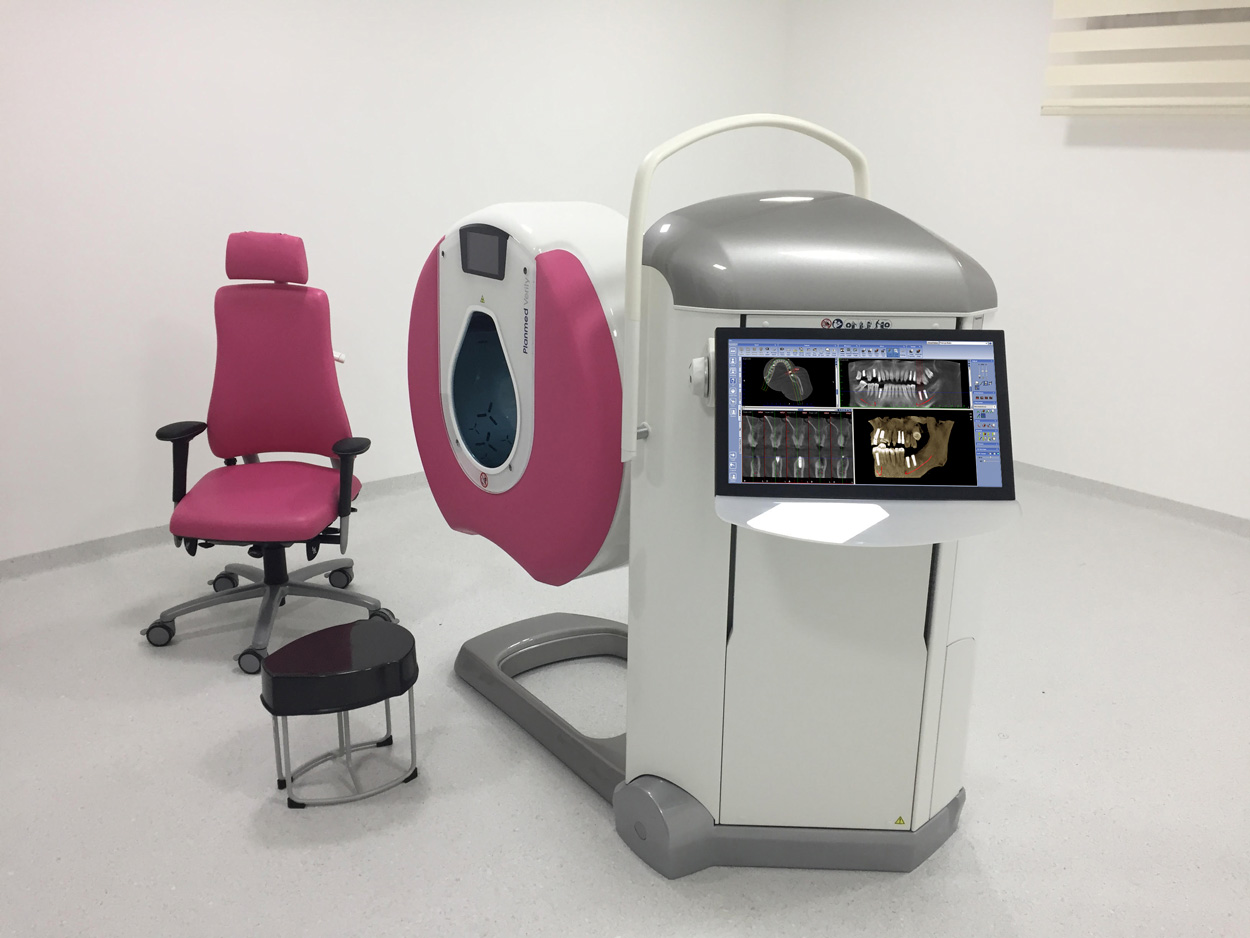
3D CBCT Scanner Planmeca Promed
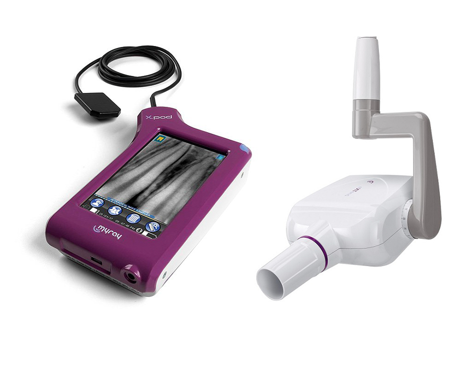
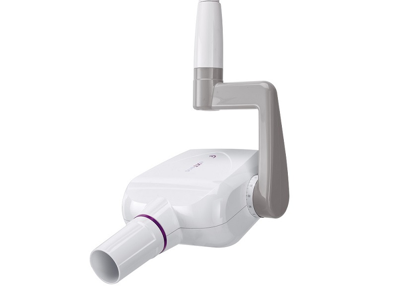
Intraoral Xray
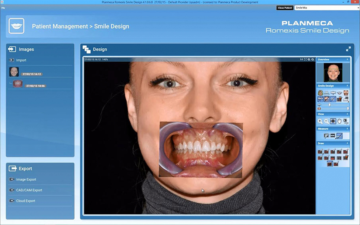
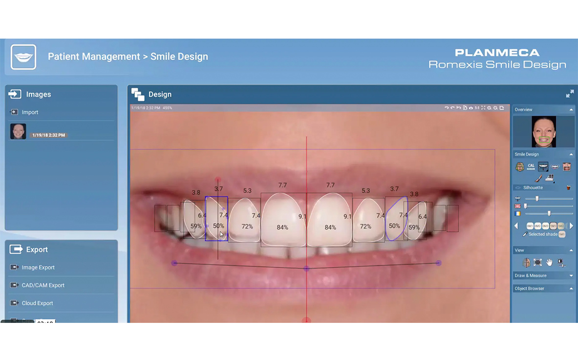
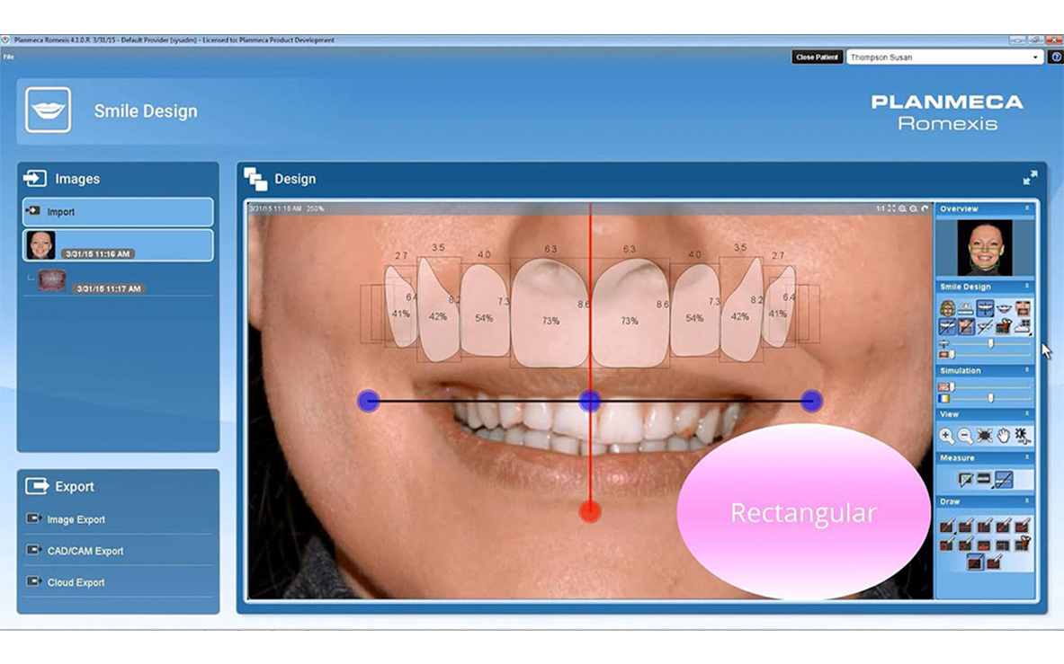
Smile design
IMPLANTOLOGY
Superior life-long solution
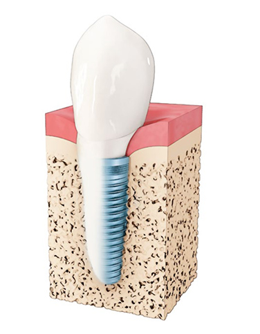
Unlike prosthesis , which entirely rests upon the gums – causing the vertical loss of underlying bone, or dental bridges – that require preparation and inclusion of the adjacent teeth, dental implants are introduced into the jaw bone – thereby simulating the natural function of the tooth root , which is to support a single tooth crown.
WHY YOU SHOULDN’T IGNORE THE TOOTH LOSS:
Emotional health and self-esteem:
– Reduced self esteem
– Reduced social interaction
– Increased risk of emotional disorders such as depression, anxiety, etc.
Self-esteem
- Before treatment 70%
- Implant placement 80%
- Final teeth 90%
Kolinski et al. 2013, De Rouck et al. 2009
– Limiting the diet ;
– Difficulty chewing ;
– Causes the movement of adjacent teeth, which can produce a change in normal bite and consequently pain in the muscles and joints of the jaw.
Physical appearance:
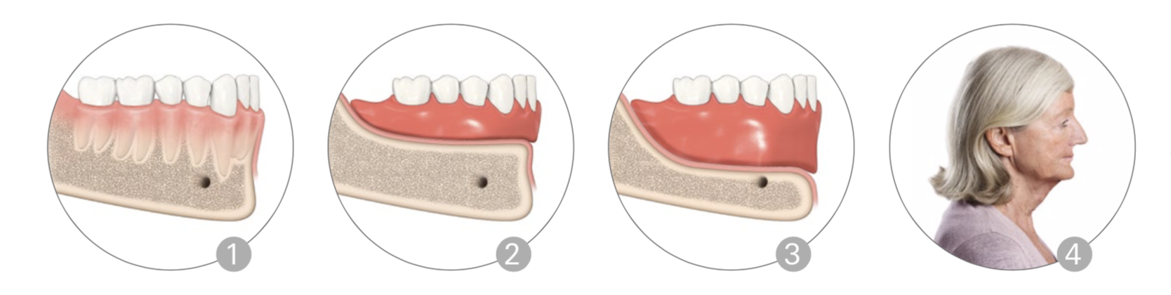
– Loss of teeth leads to the loss of jaw bone volume ;
– The loss of jaw bone volume contributes to the structural change of facial features.
Esthetics
- Before treatment 60%
- Implant placement 75%
- Final teeth 85%
Speech
- Before treatment 80%
- Implant placement 85%
- Final teeth 95%
Function
- Before treatment 60%
- Implant placement 65%
- Final teeth 95%
Sense
- Before treatment 65%
- Implant placement 75%
- Final teeth 90%
Kolinski et al. 2013, De Rouck et al. 2009
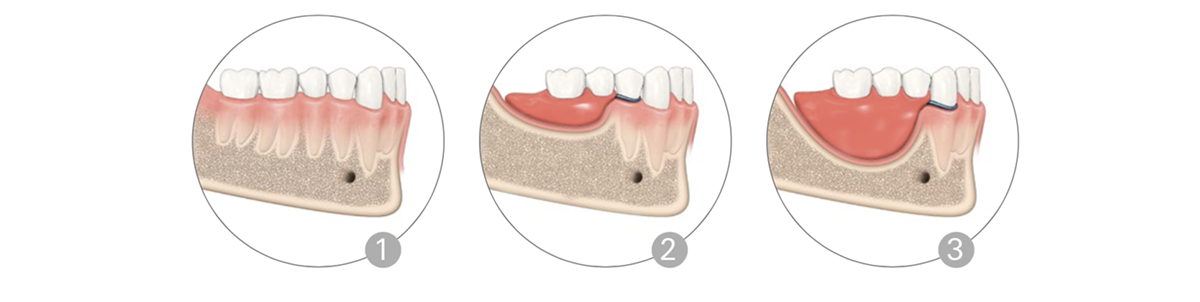
How does the lack of teeth affect the bone health :
– The absence of a single tooth leads to the reduction in volume of the surrounding bone. .

IMPLANT TREATMENT
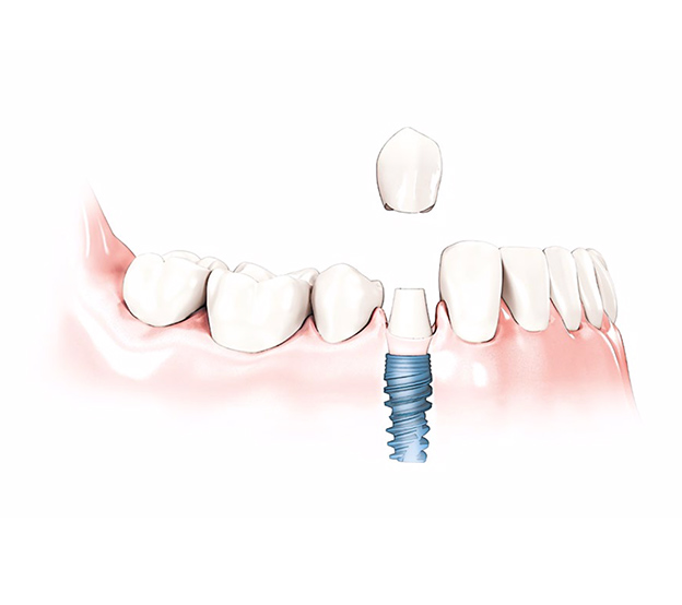
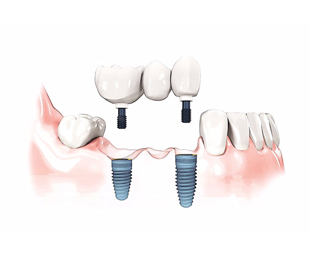
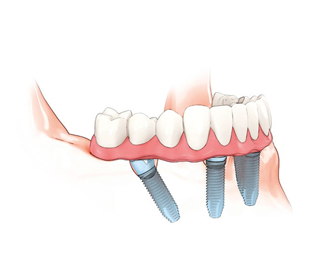
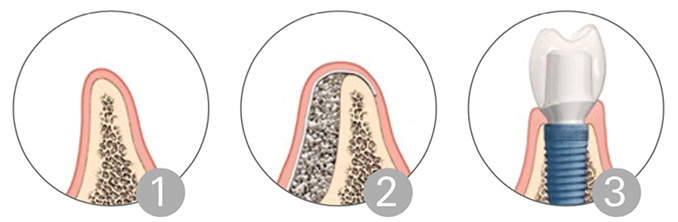
Every lost tooth may be replaced with an implant. Alternatively, fewer number of implants may support a bridge restoration in order to replace all the missing teeth.
This treatment includes the placement of 4 implants which support the fixed prosthesis, which solves the problem of missing teeth in the entire jaw whilst avoiding the problem of mobility often present with removable prosthesis.
This treatment includes the placement of 4 implants which support the fixed prosthesis, which solves the problem of missing teeth in the entire jaw whilst avoiding the problem of mobility often present with removable prosthesis.
Your personal treatment plan
Consultation and examination:
Treatment planning
Minimally invasive approach
There are two different treatment methods in implantology known as Immediate Loading and Delayed Loading.
The immediate loading technique enables the placement of dental crown on the implant, straight after the implant placement. This technique is indicated for the patients that have the optimal bone quality, and therefore sufficient primary stability of an implant. This conditions enable us to immediately “load” the implant, and complete the entire treatment in a single visit.
However, if a patient does not have an optimal bone quality (which is often the case with elderly patients), the preferred technique is delayed loading. This technique consists of implant placement followed by a waiting period of 6 months – a time necessary for hard and soft tissues around implant to mature and reach a desired stability (process known as osseointegration). Once the ‘’bond’’ between the bone and an implant is achieved, the improved stability will enable the placement of dental crown upon the implant.
Placing an implant – which entirely replaces the missing tooth, we restore the normal function and optimal esthetics, while preserving the health of the bone, gums and adjacent teeth.
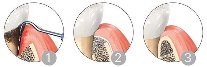
Bone augmentation
Post extraction socket preservation
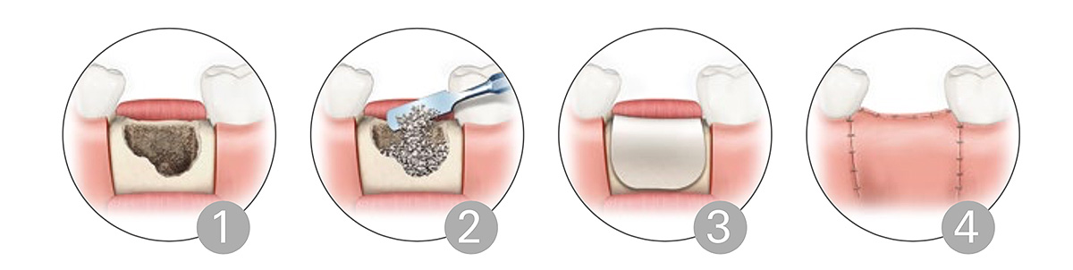
2.) Using bone grafting material to fill the bone defect after extraction for a purpose of preventing bone loss (Geistlich Bio-Oss)
3.) Using the membrane (Geistlich Bio-Gide or PRF membrane) in order to fix the bone graft.
4.) The Suturing the gums over the bone graft, while preparing for the placement of a definitive crown.
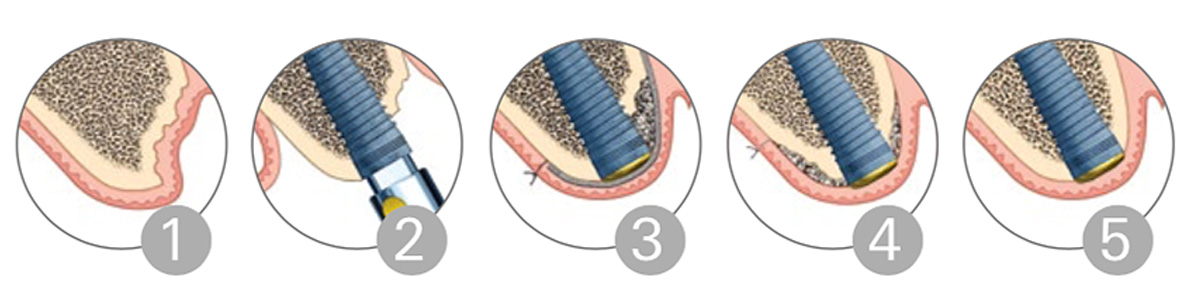
2.) Implant placement
3.) Placement of bone graft in the implant surrounding for the purpose of obtaining the optimal stability and maintaining the natural bone volume. (Geistlich Bio-Oss)
4-5.) Maturation of the bone graft and total osseointegration (bond formation between bone and implant), resulting in optimal function and aesthetics.
SINUS LIFT
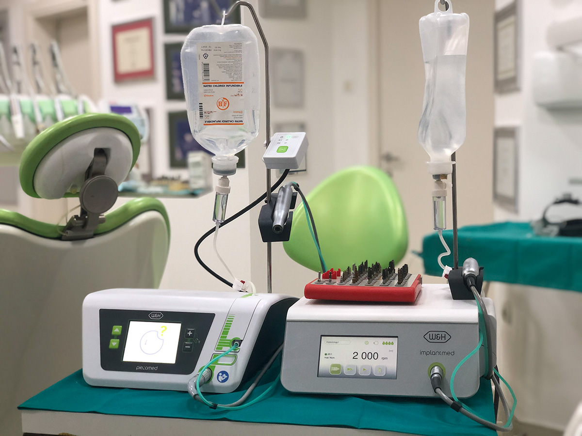
Sinus lift procedure is preformed using the advanced W&H Piezo Implant system, which increases the safety of the procedure and prevents the possibility of injuring the delicate structures in the surrounding of the maxillary sinus.
Through elevation of the sinus membrane, which represents the floor of maxillary sinus, and subsequent placement of bone graft support, we can create optimal vertical and horizontal dimensions necessary for the implant placement. Implant can be placed during the sinus lift procedure, or several months after the bone graft maturation – depending on the indication.
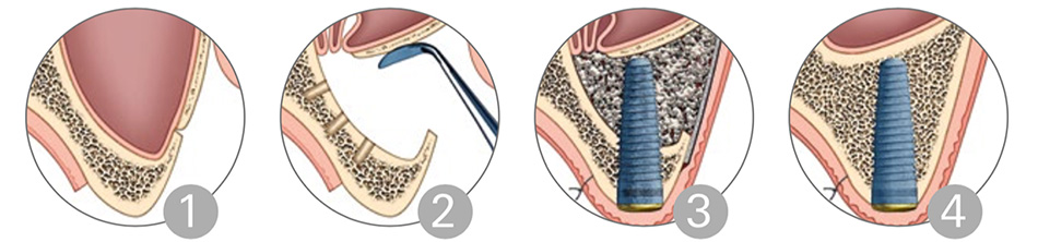
2.) Elevating the maxillary sinus floor.
3.) Bone Graft placement (Geistlich Bio-Oss) followed by the implant placement in the location of an elevated sinus floor.
4.) Completed regeneration of bone volume and implant osseointegration.
HORIZONTAL AND VERTICAL BONE AUGMENTATION
Horizontal and vertical bone augmentation is used in cases where the volume of bone in lower jaw is insufficient for implant placement.
Through the bone graft placement (Geistlich Bio-Oss), we can obtain the necessary dimensions for implant placement, either at the same time , or several months after the bone graft maturation – depending of an indication.
2.) Horizontal and vertical augmentation of the bone using the bone grafting material (Geistlich Bio-Oss)
3.) Implant placement after obtaining adequate bone dimension
GUIDED TISSUE REGENERATION (GTR)
PARTNERS:


PROSTHODONTICS
Achieving desired aesthetic outcomes.
Prosthetic rehabilitation can solve the problems of partial or total absence of teeth, irregular bite, or aesthetic concerns.
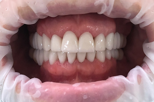
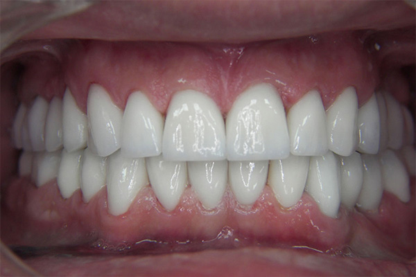
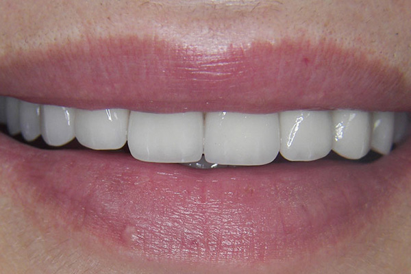
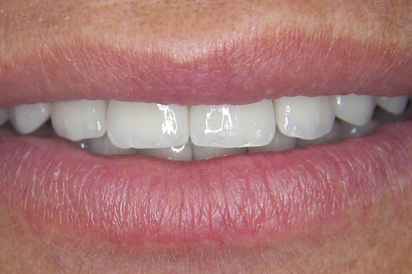
SOLUTIONS FOR TEETH REHABILITATION:
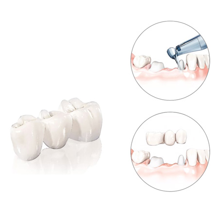
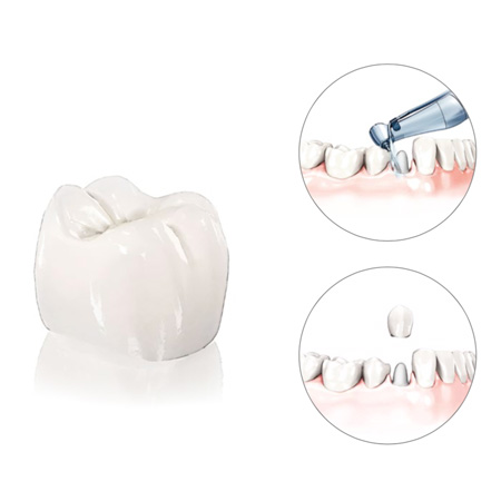
Dental crown:
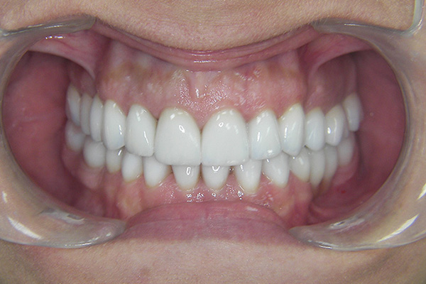
YOUR PERSONAL TREATMENT PLAN:
An indispensable part of the first appointment is the photo session – which helps us identify the main aesthetic concerns. Through the analysis and discussion, patient and dentist work together to decide which aspects of patients smile could be modified. At this stage, it is also possible to preform the Art Smile Design technique to simulate possible future outcomes, before the start of the treatment. During this consult, you will be informed about the different types of materials and techniques that are indicated for use in your case.
The second phase of treatment consists of prosthetic tooth preparation and impression taking. These impressions are used for the production of definitive crowns at our dental laboratory. After the impression taking, patient receives temporary crowns In order to preserve the normal function, appearance and soft tissue level, while waiting for the definitive crowns to be produced.
ORAL SURGERY AND PERIODONTOLOGY
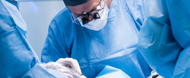
Our dental clinic can provide a wide spectrum of oral surgery treatments, both using local anaesthetics and, where indicated, intravenous sedation controlled by the anaesthesiologist.
- Extraction of impacted teeth – is one of the most common oral surgical procedures. The most common group of teeth presenting the lack of eruption or partial eruption are wisdom teeth – which can compromise the health of adjacent teeth due to their pathologic position.
- Apicotomy and cystectomy – surgical treatment of periapical lesions (lesions on the top of the root) .
- Ortho-surgical treatments – involving orthodontic extrusion of un-erupted teeth and their timely relocation to a proper position.
VRHBMP-2 (RECOMBINANT BONE MORPHOGENETIC PROTEIN-2) AND PDGF
(PLATELET-DERIVED GROWTH FACTOR)
PDGF
PRP & PRF
PRP & PRF are abbreviations for “platelet rich plasma/ platelet rich fibrin”. They can be harvested from patients own blood, and can be used in sinus lift surgery, alveolar ridge reconstruction, socket preservation, apicotomy and cystectomy. It can be used as plasma addition to the artificial bone graft, functioning as a mediator of growth factor release and stimulant for the angiogenesis process – therefore aiding the preservation of artificial bone graft volume. When used as fibrin, it can shorten the post operative healing time, and reduce the occurrence of bruising and haemorrhage.
PARODONTOLOGY
Prevention first
Periodontal disease is one of the leading causes of morbidity associated with oral cavity, which is particularly prevalent among groups of elderly and at risk patients. The disease causes resorption of tooth supporting structures leading to the compromised tooth stability, and ultimately – loss of teeth.
Periodontal disease is caused by the accumulation of dental plaque which eventually converts into harder mineralized dental calculus. To prevent transition from dental plaque to calculus, it is necessary to practice dental hygiene every day, three times a day.
It is also recommended to visit your dentist once in 6 months for a regular dental hygiene treatment.
Treatment methods for periodontitis can be either conservative or surgical:
Surgical treatment is applied in cases where progressive bone loss has occurred, and thus rendered other more conservative treatment options inadequate. Surgical treatment can include different forms of gingivoplasty and gingivectomy (surgical resection of gums).
Timely discovery of periodontal disease followed by an indicated treatment plan is of a paramount importance for maintaining the healthy state of oral cavity. Delaying the diagnosis and treatment may result in progressive development of disease, which worsens the diagnosis as the damaged tissues can no longer regenerate.
ORAL SURGERY AND PARODONTOLOGY
Minimally invasive surgery
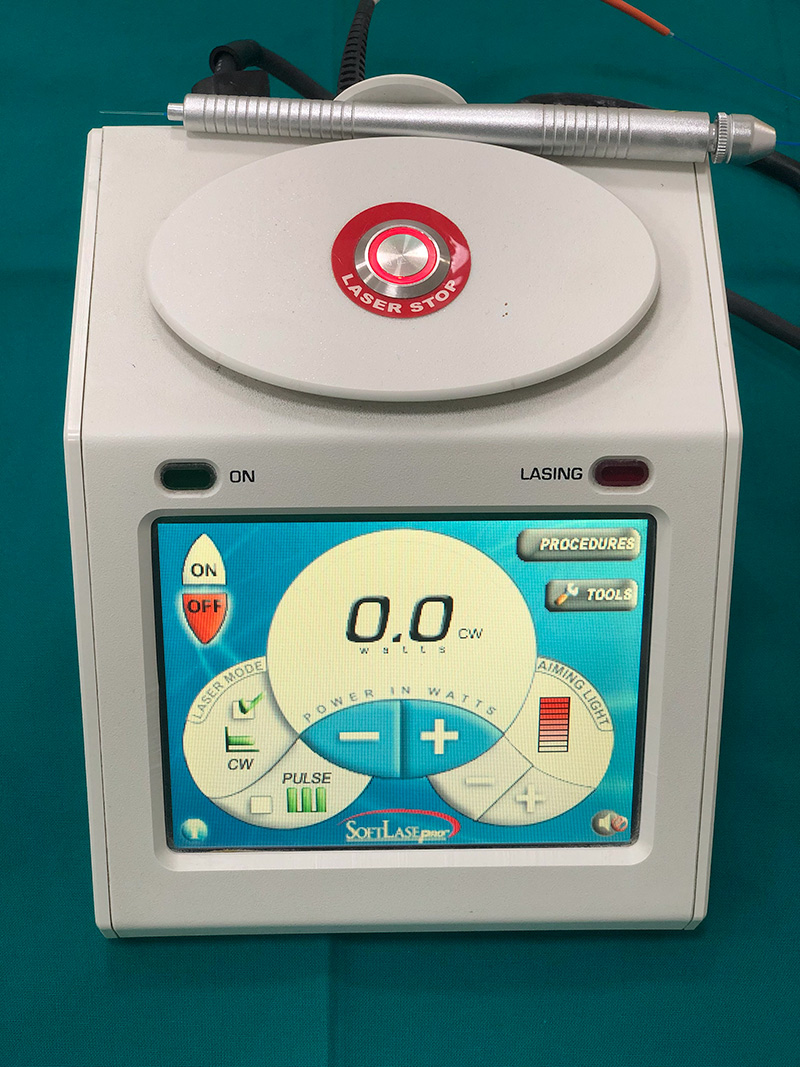
Minimally invasive surgical procedures like gingivectomy, tissue proliferation, biopsy, clinical crown lengthening, frenulum removal, apthous ulceration treatment, abscess incision and drainage, as well as the implant debridement can be preformed painlessly with the use of laser.
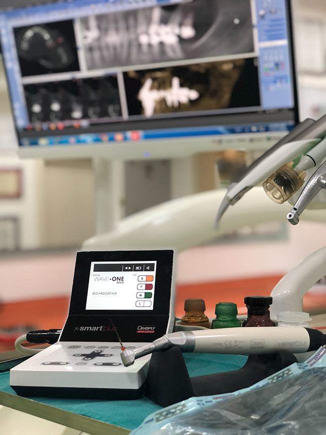
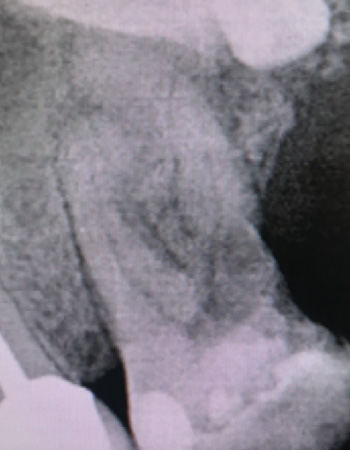
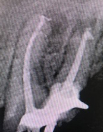
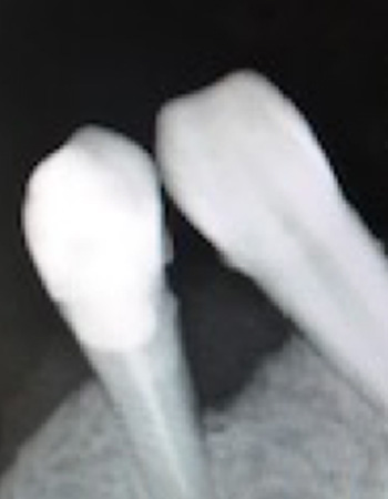
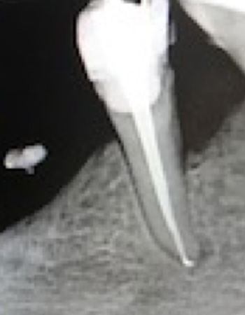
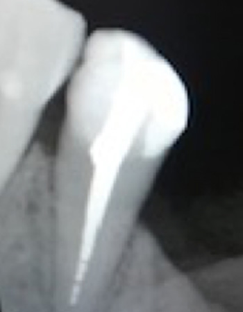
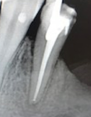
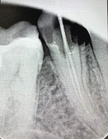
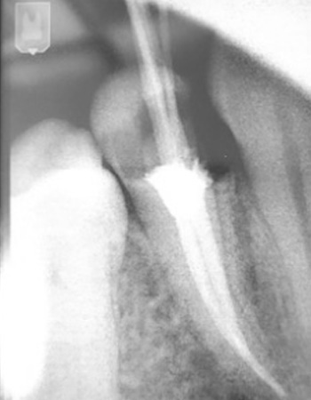
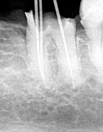
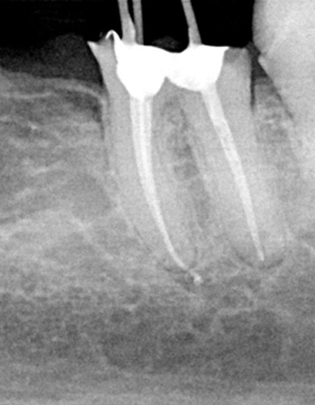
Endodontics
Treating the root cause

Endodontics is a branch of dentistry concerned with diagnosing and treating conditions of dental pulp and structures associated with a tooth root.
Endodontic treatment may be indicated in cases where:
- Patient feels pain upon exposure to an extreme temperature (e.g. while consuming cold or hot beverages)
- Pain during the consumption of sweet food
- Pain upon applying pressure to the tooth in question (e.g. when biting )
Consult and examination:
TREATMENT PLANNING:
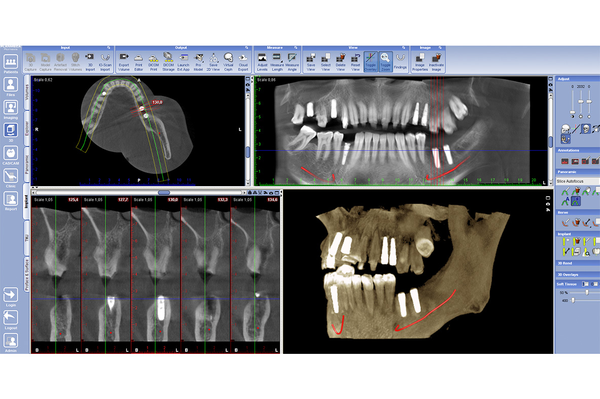
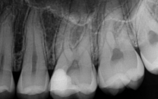
Software analysis of an Xray can be preformed in order to measure the dimensions of the root canal – a key feature of interest in root canal treatment.
MINIMALLY INVASIVE APPROACH:
Before / After
PAEDIATRIC AND PREVENTIVE DENTISTRY
Treating the most important smiles
Good hygiene maintenance, coupled with balanced diet are main prerequisites for caries prevention. The most common indicator of caries presence is improper oral hygiene.
This is precisely why it is important to carefully select the good quality tools for oral hygiene maintenance:
• Tooth brush;
• Tooth paste;
• Dental floss;
• Dental rinse .
Regular dental checkups along with preventive measures for tooth protection can help us avoid the occurrence of different pathologies of teeth and associated structures.
Preventive measures for tooth protection are : fluoridation of teeth, fissure sealing, conservative treatments of primary and secondary dentition.
FISSURE SEALING
The first permanent teeth that erupt at the age of six are molar teeth, which have an important role of forming a proper bite in the early age. This period of transition from primary to permanent dentition is often marked by the lack of proper oral hygiene. This is why fissure sealing is one of the key treatments that should be preformed at this time .
CONSERVATIVE TREATMENT OF PRIMARY AND PERMANENT DENTITION
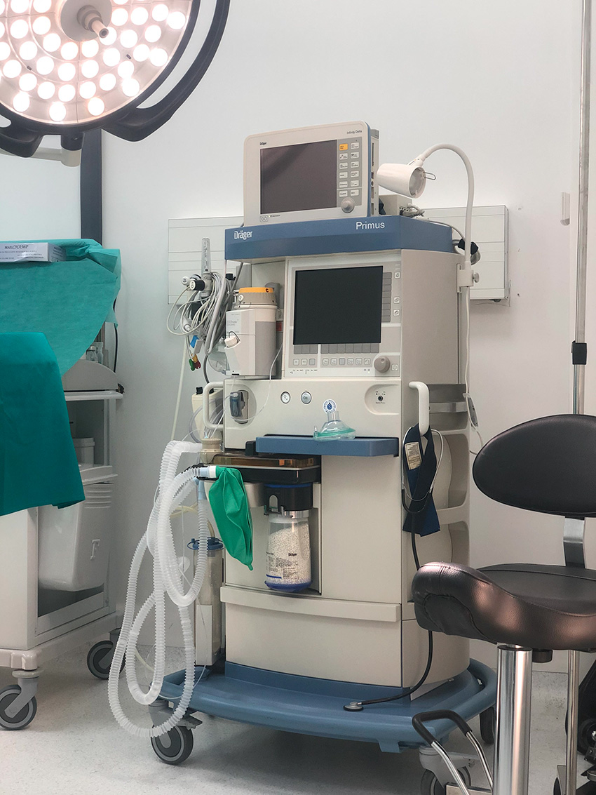
SEDATION IN DENTISTRY
Anxiety-free dental experience
Due to the worsening condition, it is often necessary to preform complex treatments that require several appointments.
Sedation in dentistry enables us to preform complex treatments in a single appointment, while minimising the anxiety and fear experienced by the patient.
Analgo-sedation is a state of reduced consciousness, which helps patient stay calm and relaxed throughout the treatment. Our team of anaesthesiologists will administer the medication before the treatment, which starts only after the patient falls asleep. Patient will remain calm, with no experience of pain throughout the treatment, and will eventually wake up with no recollection of the procedure. This type of treatment is indicated for all types of complicated procedures that last more than 20 minutes. It can be used during comprehensive implant and prosthetic rehabilitations for all the patients that experience pronounced fear and anxiety.
Dental tourism
For more than three decades, international patients have recognised dental clinic of Specialized hospital Ars Medica as the center for high quality dental treatments in implantology, prosthodontics and aesthetic dentistry.
Therefore, a special part of the clinic is dedicated exclusively to the patients coming from abroad, either for a short business trip, or a holiday. Despite the short stay, patients have the opportunity to get comprehensive dental treatment, starting with complete diagnosis and concluding with the comprehensive rehabilitation of wide spectrum of functional and aesthetic problems.
Patients can stay in one of the hospitals luxury apartments during the course of the treatment.

Contact form
SERVICES OFFERED TO INTERNATIONAL PATIENTS INCLUDE:
Free transport from the airport in Podgorica or Tivat to the clinic
Free transport from the clinic to the airport in Podgorica or Tivat.
Free accommodation in one of our luxury hospital apartments.
Assistance during reservation of hotels or other types of accommodation outside the clinic.
Assistance during organisation of touristic visits and tours.